 This presentation will focus on recent advances in modeling of the human placenta in the lab, including development of placental stem cells, organoids, and “placenta-on-a-chip.” She will describe why the human placenta remains the least understood of all human organs, including a discussion of ethical dilemmas facing researchers in this field as well as ways to overcome these barriers
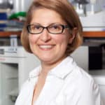
Dr. Mana Parast is the Director of Perinatal Pathology at the University of California San Diego, and a physician-scientist leading a research program focused on placental development. Specifically, her research has focused on use of stem cells to develop “disease-in-a-dish” models for studying placenta-based disorders of pregnancy, including preterm birth and pregnancy loss. Most recently, in collaboration with her colleagues in obstetrics/gynecology and neonatology, she has established the Center for Perinatal Discovery at UC San Diego, with the goal of bringing together researchers of diverse backgrounds to solve problems related to perinatal health.
Dr. Parast has disclosed that she does not have any real or perceived conflicts of interest in making this presentation.
Angela Rogers: Hi, my name is Angela Rogers, and in honor of my stillborn son, Taylor William, born October 4th, 2002, I’d like to take this opportunity to present to you, Dr. Mana Parast. She is the Director of Perinatal Pathology at the University of California San Diego, and a physician-scientist leading a research program focused on placental development. She holds a BS in Microbiology from the University of Rochester and an MD/PhD from the University of Virginia.
Dr. Parast’s research has focused on the use of stem cells to develop disease-in-a-dish models for studying placenta-based disorders of pregnancy, including preterm birth and pregnancy loss. Most recently, in collaboration with her colleagues in obstetrics/gynecology and neonatology, she has established the Center for Perinatal Discovery at UC San Diego, with the goal of bringing together researchers of diverse backgrounds to solve problems related to perinatal health.
Dr. Parast’s presentation is titled, Modeling the Human Placenta in the Research Laboratory. Welcome, Dr. Parast.
Dr. Mana Parast: Hi. My name is Mana Parast, and I’m the Director of Perinatal Pathology and the Co-director of the Center for Perinatal Discovery here at the University of California San Diego. I’m really honored to have been invited by the Star Legacy Foundation to give a talk at the annual meeting. Today I will be talking to you about how we model the human placenta in our research laboratory.
The human placenta is thought of as the most poorly-understood organ, and there are many reasons for that. But one of the main reasons is that it is also a highly evolutionarily divergent organ. What does that mean? That generally means that of the different animal models that are out there for study of various human diseases, and in particular mouse, which is used most often in the research laboratory, the human placenta is very different from those other placental mammals. The placentas of those other animals.
For example, for the mouse, not only is the structure of the placenta different, but the cell types are very different and also pregnancy associated diseases that spontaneously develop in humans, including preeclampsia, do not spontaneously arise in the mouse. While certain aspects of disease can be modeled in these research animal models, there are a lot of unique things about the human placenta, which require specifically using human placental cells in the dish.
Generally speaking, at least in the US, the human placenta is considered discarded tissue. Except when there is a pregnancy complication for which the placenta is sent to pathology for examination, the tissue is otherwise discarded at delivery. Now, the bulk of the placenta is actually derived from this outer layer of the human embryo that’s called the trophectoderm. While the inner cells over here called the inner cell mass give rise to the human embryo or the fetus, it’s these outer cells that predominantly contribute to the placenta. In fact, the placenta is an embryo or fetal derive tissue, but it’s distinct from the embryo and fetus.
There are, of course, maternal components to the placenta. This includes maternal blood, which flows into the placenta and helps to nourish the placenta as well as the lining of the uterus called decidua, which provides a manner for the placenta to be attached to the uterine wall.
In early gestation, the human placenta starts to develop like a young sapling. If you consider this fetal surface here to be the roots, you can see that the branches and the leaves are growing out of the roots and forming the functional unit of the placenta called villi or singular villus. These villi grow out towards the maternal surface and there are portions of them that are floating in maternal blood and are referred to as floating villi. These are the structures that are closer to the fetal surface. Then as they approach the maternal surface, they attach to the decidua, the uterine lining, and these villi in particular are referred to as anchoring villi. I’m going to introduce you to these two different functional parts of the placenta separately.
First, the floating villi. These are predominantly for nutrient and gas exchange. What you’re seeing here is a cartoon of a single villus, which is kind of like a finger-like projection in cross section. This villus is floating in maternal bloods. It’s based on maternal blood on the outside. It is lined by two layers of placental cells. The inner layer is called cytotrophoblast or CTB. The outer layer is composed of these cytotrophoblasts that have fused to form a multinucleated layer of cells called syncytiotrophoblast.
Then inside the villi are other cell types that basically surround these branches of umbilical vessels, and so these are fetal blood spaces that are shown here to be filled with fetal red blood cells. Nutrient and gas exchange between the fetal blood and maternal blood have to take place across this interface.
Towards the maternal surface, there are these anchoring villi, that as I mentioned, help attach the placental unit to the uterus and at this interface, these cytotrophoblasts stem cells, instead of making syncytiotrophoblast, they instead form these invasive cells that are referred to as extravillous trophoblasts, and these extravillous trophoblasts or EVT help attach the placenta to the uterine wall.
Now, a subset of these cells also do something really cool, which is that they remodel maternal vessels in the uterine wall. Before maternal vessels get remodeled, they’re sort of small. You can see that the lumen that would carry blood to the uterus is seen here, and that’s pretty small. After the trophoblast, these extravillous trophoblasts, invade and remodel the vessels, and in this process what they actually do is they replace the maternal endothelial cells, the mom’s cells that normally line the vessels, they get replaced by these extravillous trophoblasts. You can see that the difference is a very dilated vessel that can therefore carry more blood to the fetal placental unit and therefore provide the oxygen and the nutrients that the placenta and the baby need during pregnancy. These cells are really important. If they don’t function well, and they don’t get established well early in gestation, complications could arise including intrauterine growth restriction and preeclampsia.
I’ve been using this word trophoblasts a lot. Trophoblasts are the main functional cells in the placenta. The meaning of the word trophoblast, the word trophoblast comes from the Latin root tropho, which means to feed. In fact, in the placenta, there are these stem cells, early on in gestation, called trophoblast stem cells.
These are part of the cytotrophoblast layer that I described earlier, and these cells are stem cells which by definition means that they can self renew. They can make more of themselves, and they can differentiate or develop into both the extravillous trophoblasts that I described, which are invasive cells that anchor the placenta to the uterine wall, and also help in terms of having a conversation, a cross talk with maternal immune cells so that the placenta doesn’t get rejected.
These cells, these trophoblast stem cells can also differentiate in the floating villi into syncytiotrophoblasts. These are the multinucleated cells that secrete HCG among other hormones, and are also involved in gas, nutrient, and waste exchange. So really in order to be able to study the human placenta, we need a model. We need a human trophoblast stem cell model that is able to self renew and make both these types of cells.
Now, if we start with a term placenta and isolate cytotrophoblast cells from a term placenta, these cells cannot make extravillous trophoblasts. The only cells they can turn into are the multinucleated syncytiotrophoblast. What’s more is that these cytotrophoblasts do not self renew, so that if you want to do an experiment with the cells, you have to basically do cytotrophoblast props from multiple different term placentas.
Now, in contrast, if you cytotrophoblast from early gestation placenta, before 10 weeks gestational age, these cells do have the ability to become both EVT, extravillous trophoblasts and STB, the syncytiotrophoblasts. Now until recently, these cells also could not be cultured continuously in the dish, so that cytotrophoblast perhaps had to be done from different placentas for each separate experiment.
However, because again, we know that these cells are able to differentiate into both of these cells, so at least the portion of them must be stem cells. One group was able to develop culture conditions, culture media with the necessary growth factors in order to be able to derive trophoblast stem cells. This work was published about three years ago. This was developed in the lab of Dr. Arima in Japan. These culture conditions were applied to early gestation placenta, and so you could derive these self-renewing stem cells.
You really only have to do one prep, and then you could have an indefinite number of cells to work with that could be frozen and thawed and experiments could be done on the same isolated cell. They could self-renew for prolonged period of time for months on end, and so it could be used for an indefinite number of experiments. Dr. Arima’s group also showed that the same cells could also be derived from human embryos. These are embryos that are leftover after patients’ IVF cycles are completed.
These cells can be grown in two different ways. Generally speaking, when we grow trophoblasts themselves or cytotrophoblasts in culture, most people grow them in a flat dish, so in two dimensions or what are called flat culture dishes. But placental tissue or cells can also be grown in three dimensions. In the past, the most popular type of this type of three-dimensional culture was placental explants. Taking small bits of placentas, and again, this could be done from either term placenta or early gestation, and these explants could be used in the dish for a few days, and of course new props had to be done each time.
The advantage of a three dimensional culture is that it gives you different types of cells, especially the explants in the correct context. The cells in relation to each other are very similar to what their environment is like in in vivo in the human body.
Now over the past few years, new methods of three-dimensional culture has been developed. The terminology that’s used for this is organoid. Organoids have been developed from many different organ types, including intestinal organoids, but recently this has also been developed for the placenta or what’s called trophoblast organoids have been developed. This is from work from a group in Cambridge that was published about the same time, about three years ago, where, again, cells from first trimester placenta were able to be organized in an organoid form and cultured, again, indefinitely just like human trophoblast stem cells in the dish.
It was also found at the same time that while you could make organoids directly from this tissue, you could also first isolate the trophoblast stem cells, the way Dr. Arima’s group had developed, and then take those cells that were normally growing in two dimensions and culture them in three dimensions in such a way that they grow as an organoid. This can be done both ways.
Another relatively new method of three-dimensional culture is what’s called organ-on-a-chip, and again has been done for multiple organs recently, and it’s also been done for the placenta. There are multiple groups that are working on this, including our group here at UC San Diego. What this does is in particular this method is aimed to model the nutrient and gas exchange interface.
This is the villas interface that has both trophoblasts and fetal endothelial cells that separate the barrier between fetal blood and maternal blood where gas and nutrient exchange must occur. This basically takes advantage of cells that are derived or cell lines that are available that can be organized within particular compartments and such that there can be two layers of cells that are organized exactly as they would be in vivo. Then the exchange across the interface for whether it’s drugs or glucose can be tested across this barrier.
This is another picture from another group’s placenta-on-a-chip where endothelial cells are shown in green and the trophoblast cells in red. Again, this is trying to show that you can form a very nice barrier of both these trophoblasts and endothelial cells and use them to transfer various drugs or nutrients across the surface.
There are a lot of advantages of three-dimensional culture of these placental cells. As I mentioned, for organoids, you can actually see better differentiation or development of specific cell types that more closely approaches what happens in vivo in the body. With placenta-on-a-chip, the ability to evaluate how nutrients or various drugs are transferred across this maternal fetal interface is really important because in two dimensions it’s difficult to form a very good trophoblast barrier, but under conditions of flow and in the right context in combining the various types of cells, this can be better modeled in this manner as placenta-on-a-chip.
What are some potential sources of hTSC? The main source for these cells is from elective surgical terminations of pregnancy. NIH guidelines require that consent be obtained for tissue donation, and that this consent is obtained remote from patient consent for the procedure itself. In other words, the patient first consents for the procedure and has decided to terminate her pregnancy before she’s approached to donate the tissue. Specifically, surgical procedures cannot be altered for research purposes. The derivation efficiency from such samples is greater than 90%.
As I mentioned, human TS cells can also be derived from embryos, from intact embryos. Again, these are usually excess embryos that are donated by patients once they’ve completed their IVF cycles. Again, consent is required and efficiency of derivation is also high for this source.
These cells can also be derived from cells following miscarriage, and again, NIH requirements require consent for this. However, because by definition, the tissue is not viable, the efficiency of derivation of such cells is significantly lower for this material.
Now, there are, of course, as you might imagine, limitations from such sources. One limitation is that in fact work or research with human embryos in the US is not funded by the NIH. Placental tissues from elective termination of pregnancy require additional justification for use by NIH. Of course, there are ethical objections to use of either of these tissues as sources for derivation of human TS cells.
For the past few years, researchers have also looked for alternatives to use of such tissues for developing models for studying the human placenta. I’m going to talk about one of these really promising alternatives. To do that, I’m just going to go back a little bit and first talk about another cell culture model, and this is human embryonic stem cells.
Human embryonic stem cells were first derived from embryos in the late 1990s and early 2000s. By definition, these were cells that could give rise to any embryonic or a fetal cell type, including cardiac muscle, lung cells, skin cells, neurons, so cells from the brain. When this technology was originally developed, it truly revolutionized the study of human biology, especially early human development.
However, again, in order to derive such cells, human embryos were needed, and so again, there were ethical objections to use of human embryos for this kind of research. Shortly thereafter about 2007, the first publication on induced pluripotent stem cells came out, in which basically researchers had developed a method to turn any adult cell into a cell that pretty much acted like a human embryonic stem cell.
In this protocol, what you need is basically a cell type that would come from already developed tissue and adult cell, but it needed to be proliferative. For that reason, a lot of times skin cells were used. Skin cells are proliferative, and of course they’re easy to access. You could start with skin cells and introduce three or four, what were called iPS reprogramming factors. These are genes that were introduced for just a short period of time into these proliferative cells.
After about three to four weeks of these genes being expressed, in the tissue culture dish, you would see colonies of cells arising, which for all practical purposes looked and behaved like embryonic stem cells. One could basically apply the same protocols and differentiate these iPS cells into cardiac muscle, again, lung cells, skin cells, brain cells, any type of embryonic or fetal cell or tissue types.
Now, around the same time that this technology was coming about, multiple groups were finding out that iPS cells, human iPS cells could also be converted not just to embryonic and fetal cells, but also into placental cells or trophoblast. Multiple different methods for doing this have been developed including one in our lab. The first thing that basically has to be done is the cells have to be put into what’s called trophoblast induction media. For a few days, the cells are exposed to growth factors that push the cells toward a cytotrophoblast like cell. Then basically the human TS media could be applied to these cells. After a few passages in culture, you basically end up with these colonies of cells that again, for all practical purposes, look and behave like human trophoblasts themselves.
Based on this, we are using this method in order to develop models for studying multiple different types of human placental disease. What our lab has done is that we start with umbilical cord or cells from the amnion. These are cells in the placenta that are very clearly derived from the fetal portion of the placenta. So they have the same genetic material as the baby. We introduce these reprogramming factors into these cells. After a few weeks, we get iPS cells. Then using the protocol that we’ve developed in our group, we convert these cells to trophoblast stem cells, and now we have trophoblast stem cells that basically can tell the story of that particular placenta in that particular pregnancy.
This is very exciting for us. We can actually start, again, like I said, with any placenta at delivery and generate iPS models, which means that we can generate iPS models for the study of preeclampsia, preterm birth, gestational diabetes, and placenta accreta spectrum. This latter disease is a very horrific disease whereby the placenta doesn’t separate from the underlying uterus. In fact it’s understood that this might at least partially be because the trophoblast are a bit too invasive.
We can also use iPS cells to study gene environment interactions. Like I mentioned, the iPS cells that are developed from the placenta share the same genetic material as the baby. You can basically expose those cells. You can turn them into placental cells and then expose them to various environmental conditions that we know are associated with pregnancy complications.
For example, hypoxia or low oxygen, inflammation, which is very highly associated with different kinds of preterm birth but also with preeclampsia can expose them to pathogens and evaluate the responses of different iPS cells from different placentas to these alterations in the environment.
iPSC-derived trophoblast can also be incorporated into placental organoids and to placenta-on-a-chip, so that this way you can, for example, understand the behavior of preeclampsia associated iPS cell in the context of three-dimensional models such as the placenta-on-a-chip. For example you could ask if one particular iPS cell when turned into trophoblast is able to respond to various– it can do a better job, for example, in nutrient transfer than another iPS cell from a different placenta, and differences in these iPS cells could potentially explain why there was fetal growth restriction that complicated one particular pregnancy.
iPS cells can also be made from both mom’s cells and placental cells at the same time. You can take the placental iPS cells and turn them into trophoblasts. You can take mom’s iPS cells and turn them into various cells resembling immune cells or decidual cells, the cells of the uterine lining. Then you can put these cells into the same three-dimensional culture and see how these cells interact or crosstalk and model what would have taken place, what you would see at the maternal fetal interface which is basically an area that you don’t have access to in an ongoing pregnancy. Again, these iPS cells have tremendous potential which can be used in order to understand pregnancy complications that otherwise cannot be modeled using animal models or other methods.
I hope I have gotten you very excited about what’s happening in the study of the human placenta, what has happened the past few years, and we’re really, I feel, at a forefront of a new era of research into human pregnancy because we have all these tools in order to be able to understand both the development of this organ during normal gestation, and also be able to manipulate the system in order to study placental disease.
I invite you to check out the website for our newly established Center for Perinatal Discovery, and of course, if you have any questions, please don’t hesitate to email me. I hope you join us in participating in studies where you’re able to and help us understand pregnancy complications and really bring forth everybody’s energies, both from patients’ perspective and from researchers’ perspective together to tackle understanding of the human placenta and human placental disease. Thank you so much.
This presentation was part of the Stillbirth Summit 2021. This individual lecture will be awarded .5 hours of continuing education credit to include viewing the lecture and completing evaluation and post-test. Once received a certificate will be emailed to the address you provide in the post-test. If you did not register for the Summit WITH Continuing Education, you can purchase the continuing education by clicking here. This purchase will provide you access to all Stillbirth Summit 2021 lectures including continuing education credit. There is no charge for viewing the presentation.

To receive continuing education credit for this lecture, the participant must complete the evaluation and post-test.
Please feel free to ask questions of the presenter. We will obtain their answers/comments and provide them here as received.
Add your first comment to this post